


With the bi-directional standing weight-bearing full-length imaging system installed in Jinan in the second quarter of this year, on the afternoon of June 11, 2026, the "Bi-Directional Standing Weight-Bearing Full-Length Imaging System - Orthopedics and Radiology Academic Exchange Conference" hosted by the Second Qilu Hospital of Shandong University was successfully held in Room 4 on the fourth floor of the Qiuzhen Building. The conference was moderated by Professor Zhong Hai of the Department of Radiology, and President Feng Shiqing delivered the opening remarks.
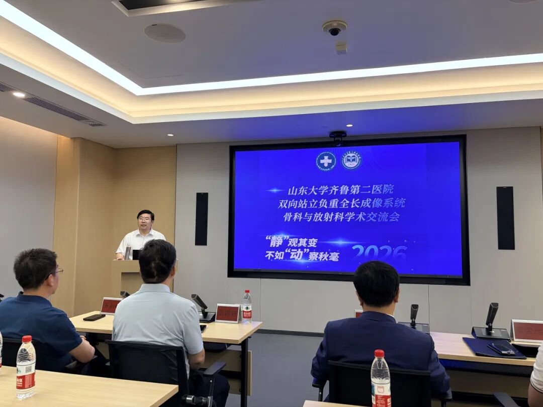
Professor Zhong Hai moderates the meeting

President Feng Shiqing delivers opening remarks
The conference brought together many top experts and scholars in the fields of orthopedics and radiology from within the hospital and other provinces, conducting in-depth discussions aroundthe bi-directional standing weight-bearing full-length imaging technologyits clinical application value, contributing rich academic wisdom to advancing orthopedic diagnosis and treatment toward precision, functionalization and digitalization.

Traditional orthopedic imaging assessment relies mostly on the basic morphological data provided by two-dimensional X-ray, non-weight-bearing CT or conventional MRI. However, for many foot and ankle, knee, hip and lower limb deformity diseases, the patient's real pain, deformity, instability and functional impairment often occur in real physiological states such as standing, walking and weight-bearing.
Observing changes in the "static" is not as good as detecting subtle differences in the "dynamic". The bi-directional standing weight-bearing full-length imaging system focused on at this conference is a powerful response to this clinical pain point. The system not only achieves weight-bearing full-length imaging of the whole spine and whole lower limbs, but also ensuresmore accurate and realistic slit-scan imaging results than traditional cone-beam DR technology, and also introduces, in a breakthrough manner,bi-directional synchronized dynamic imagingandfunctional assessment mode, marking that the Second Hospital of Shandong University has entered a new stage in the field of orthopedic functional imaging, and will provide more comprehensive, realistic and measurable imaging evidence for patients with scoliosis, joint diseases and lower limb deformities.

PLA General Hospital Professor Zhang Guoqiang
"Longitudinal Analysis of Knee Motion Differences Between KOA Patients and Healthy Individuals Using Multimodal Imaging"

Xi'an Honghui Hospital Professor Tang Yonggang
"Analysis of X-ray Slit Scanning and Dynamic Medical Imaging"
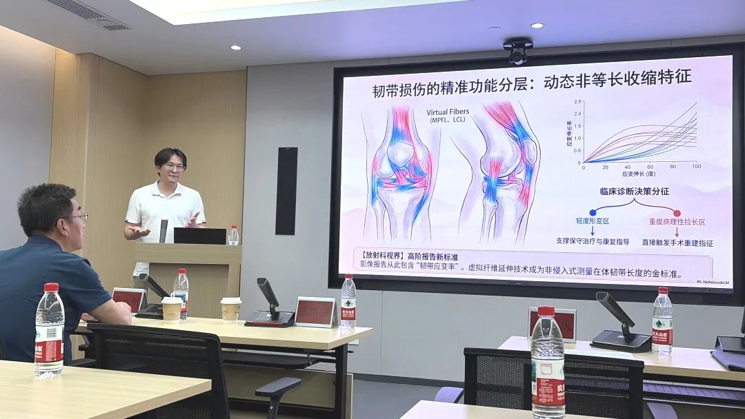
Shanghai Jiao Tong University Professor Cai Zongyuan
"From Static to Dynamic: The Clinical Value of Dual-Plane Imaging in Precision Orthopedic Medicine"

Traditional orthopedic imaging mostly uses supine or seated non-weight-bearing scanning. Once the patient lies down, the joint space, alignment and stability often "disguise" as normal; but once they stand up, walk and squat down, the real misalignment, imbalance and compensation are fully exposed.
Bi-plane synchronized imaging is precisely the key technology to uncover this "disguise". The patient is scanned in a natural standing posture, with the lower limbs or whole spine bearing real body weight; through synchronized exposure of dual orthogonal X-ray sources, the system obtains frontal and lateral orthogonal images in a single capture, completely eliminating the geometric distortion caused by projection angles and posture changes in traditional X-ray films, achieving 1:1 distortion-free reproduction.

If bi-plane synchronization solves the problem of "viewing angle and positioning", then full-length slit scanning completes the puzzle of "wholeness and distortion". Traditional X-ray often captures only one joint, but the human skeleton is a mechanical chain - if the foot and ankle are misaligned, the knee compensates; if the pelvis is tilted, the spine curves accordingly.
Full-length slit scanning technology enables patients to obtain, in a single scan under standing weight-bearing conditions, ultra-long field-of-view images from the soles of the feet to the skull base (whole lower limbs, whole spine, or full body length), while ensuring synchronized frontal and lateral imaging and 1:1 distortion-free imaging through parallel-light slit scanning combined with better imaging technology. Based on this "full-length panoramic image", doctors can complete cross-joint overall assessment of the foot-ankle-knee-hip-pelvis-spine within the same coordinate system.
This technology has achieved two practical functions:
2D AI measurement: rapidly quantifying lumbar-pelvis-hip complex parameters and identifying normal or functional abnormalities;
3D spatial reconstruction: reconstructing three-dimensional bone models from two-dimensional images and measuring parameters such as angles, distances and rotations in real coordinates, moving from "empirical speculation" to "data-based positioning".
The above technologies improve the goal of precise imaging at the static structural level, but when facing various functional problems such as dynamic ligament laxity, abnormal patellar tracking, spinal instability or restricted mobility, instantaneous joint dislocation, pinching and impingement during movement, one still has to scrutinize the details in dynamic images.
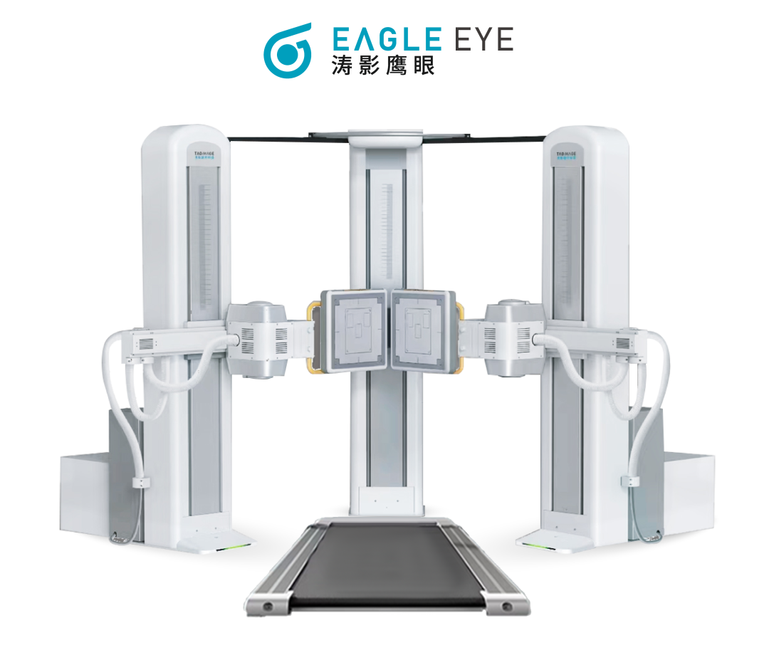
Dual-Plane Bone and Joint 4D Dynamic Imaging System

Clinical needs have never stopped at "seeing structure". What doctors truly long for is - what exactly happens to the joint at the moment the patient stands up, walks and squats down?
To this end, on the static foundation of the bi-directional standing weight-bearing full-length imaging technology, TaoImage Medical has innovatively added theDual-Plane Bone and Joint 4D Dynamic Imaging System. This system extends the assessment dimension from "space" to "time", leaping from "static anatomy" to "dynamic function", achieving the advanced application of functional imaging. It is not meant to take a clearer picture, but to answer a more fundamental question: compared with normal people, what problems in the affected part do the differences shown by the patient during movements actually reveal?
Knee Joint Dynamic Imaging
From bi-plane three-dimensional modeling to 4D dynamic motion capture, the system frees orthopedic doctors from "inferring dynamic function based on experience". Whether it is the assessment of dynamic laxity before anterior cruciate ligament reconstruction or the verification of functional recovery after patellofemoral disorder surgery, TaoImage Medical's Dual-Plane 4D Dynamic Imaging System brings functional imaging truly into daily clinical practice, letting every diagnosis and treatment decision be supported by dynamic data.

The successful holding of this academic exchange conference not only reflected the high importance that domestic orthopedic and radiology experts attach to functional imaging and dynamic assessment, but also released an important signal: future orthopedic diagnosis and treatment will pay more attention to the mechanical state of patients in real weight-bearing and movement, and emphasize the integration of static and dynamic, quantitative precision and individualized full-chain management.
For dual-plane dynamic imaging technology, this is both an inevitable direction of the academic frontier and a major opportunity for clinical implementation. TaoImage Medical will continue to focus on the real clinical needs of orthopedics and rehabilitation medicine, promote the in-depth application of the Dual-Plane 4D Dynamic Imaging System in precise imaging of whole-body joints (shoulder, elbow, hip, knee, ankle, etc.), lower limb alignment analysis, spinal disease diagnosis and treatment, and sports injury assessment, empower preoperative planning, postoperative follow-up and research translation, and jointly explore new paths of precise assessment "from static to dynamic" with experts and colleagues, helping Chinese orthopedic diagnosis and treatment reach a higher level.
Recommended Reading
Recommended Reading

Related News
-
 Product Innovation
"Sports Medicine Research" Magazine Officially to Be Launched!
2026.04.13 -
 Media Reports
[Reprint] Sports Medicine Frontier: "Breaking New Ground, Leading the Future | The Cross-Regional Chinese Artificial Ligament and Patch Research Society Officially Established"!
2026.06.01 -
 Academic Activities
Recap | Ligatech Academic Meetings, January 2026
2026.02.09